A new technology offers researchers the chance to decode the structural makeup of methane’s conversion to methanol.

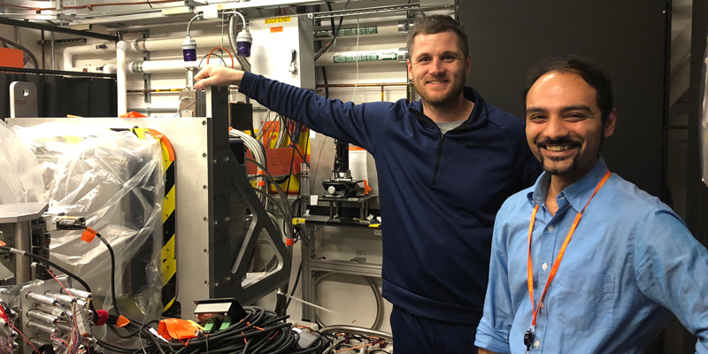
Jason Jones (pictured left) and Dr. Rahul Banerjee (pictured right) at the Stanford Linear Accelerator Center
For Jason Jones and Dr. Rahul Banerjee, finding clarity in their research is often complicated. Jones, a fourth-year Ph.D. student, and Banerjee, a postdoctoral associate, study soluble methane monooxygenases (sMMO) in Dr. John Lipscomb’s lab. sMMO is the enzyme that converts methane into methanol, but the biochemical processes underlying this conversion are not fully understood. The two recently had a chance to take advantage of a unique technology to try to see down to the electron level of what happens in the methane to methanol conversion.
“The only way we can really see what’s going on there is if we get a picture of the enzyme reaction via x-ray crystallography,” says Banerjee. “With the instrument at the Stanford Linear Accelerator Center, the idea is that you’re putting so much power into each x-ray pulse, you get a diffraction pattern, or image from the light, before there’s destruction.”
This last May and December, the two joined collaborators from around the world at the Stanford Linear Accelerator Center to use the Linac Coherent Light Source X-ray Free Electron Laser ( LCLS XFEL), one of only five such x-ray crystallography machines in the world. The two are also the first researchers from the University of Minnesota to use such an instrument.
X-ray crystallography is a process that involves using an x-ray beam to evaluate the structure of proteins. To make it work, researchers use machines that shoot x-rays at crystallized forms of substances, but this approach can have drawbacks. When researchers use traditional x-ray crystallography machines, such as synchrotrons, the x-rays themselves can damage the crystallized proteins, so they can not get an accurate depiction of the protein structure when methane gets converted to methanol.
“When you use an XFEL for x-ray crystallography, it creates this very brilliant light,” says Jones. “With that brilliance comes a lot of energy and it’s that high energy that’s being used to study systems that are sensitive to radiation damage, such as sMMO.”
That’s where the XFEL comes in. Because the instrument emits such a strong and quick x-ray laser beam compared to synchrotrons, researchers can obtain protein crystal structures before radiation damage obscures the true image. Jones and Banerjee have some preliminary results from their time in California, but the researchers will need to do additional experiments on XFEL machines for a complete picture of the enzyme they study. But they see real opportunity to learn from these results.
“We’ve focused so much on the chemistry, but there’s protein around that in this reaction and there’s so much more,” says Jones. “My hope is that some bright person would get that information and think ‘Oh, that’s what I needed to make this new synthetic catalyst, with potentially broad implications in areas like methanol production or the transportation of natural gas.”
-Lance Janssen